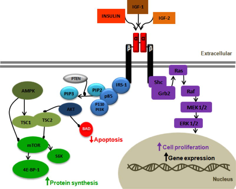
Figure 2.
Insulin-like growth factor 1 receptor (IGF-1R) signaling pathway. Binding of IGF-1 or IGF-2 or insulin to the IGF-1R α-subunit leads to autophosphorylation of β-subunit residues, which then act as docking site to insulin receptor substrates (IRS-1 to 4). IRS-1 recruits the p85 regulatory subunit of phosphatidylinositol 3-kinase (PI3K) which activates the p110 catalytic subunit, then resulting in the formation of phosphatidylinositol 3,4 phosphate (PIP2) and phosphatidylinositol 3,4,5 phosphate (PIP3). PIP3 activates Akt. Activated Akt has many substrates; in one pathway Akt inhibits apoptosis by inactivating BCL-2 antagonist of cell death (BAD), and in the second pathway Akt regulates protein synthesis by phosphorylating tuberous sclerosis complex (TSC1/2). This phosphorylation removes the inhibition of TSC1/2 from mammalian target of rapamycin (mTOR). mTOR activates the ribosomal S6 kinase (S6K) and eukaryotic initiation factor 4E-binding protein-1 (4E-BP-1), leading to protein synthesis. In the absence of cellular nutrients, AMPK can inhibit protein synthesis through mTOR inhibition, both directly and by activating the TSC1/2 complex. The tumor suppressor phosphates and tensin homolog deleted on chromosome 10 (PTEN) inhibits PI3K. The mitogen-activated protein kinase (MAPK) pathway can also be activated by IGF-1R activation. In this pathway IGF-1R activates the adaptor proteins, She and Grb2, leading to activation of Ras, Raf, MEK1/2, and ERK1/2, which results in cell proliferation.