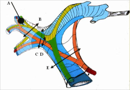
Figure 2.
Glissonian pedicle elements: portal vein (in blue), hepatic artery (in red), and the bile ducts (in green). Hepatic inflow occlusion: A) Selective occlusion of segmental portal vein by a balloon introduced under ultrasonographic guidance. The arrows show the different sites of Glissonian clamping. B) Suprahilar clamping of one sector of the right liver; C and D) hilar selective clamping to the right liver; E) total pedicular clamping (Pringle maneuver).