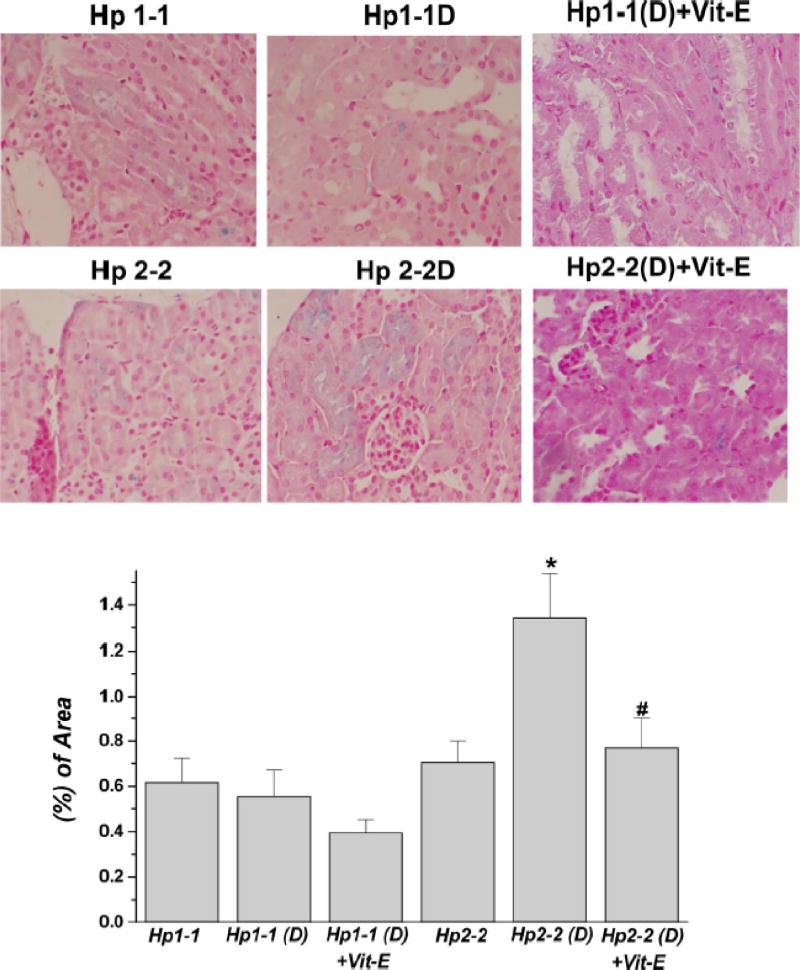
Figure 4.
Increased renal iron deposition in the proximal tubule of Hp 2-2 mice with Diabetes Melitus (DM). Perl’s iron stain was used to localize iron in paraffin-embedded kidney sections in Hp 1-1 and Hp 2-2 mice with and without DM. Arrow indicates iron-induced stain in blue (×400 magnification) located within proximal tubular cells. There was a significant increase in iron staining in the renal tissue of Hp 2-2 DM (D) vs. Hp 1-1 DM (D) and Hp 2-2 non-DM mice (P < 0.001; n = 6 animals for each group). *Hp 2-2 (D) vs. Hp2-2. # Hp 2-2 (D) + Vit-E vs. Hp 2-2 (D). Figure included with permission from American Physiological Society. (Nakhoul et al.26).