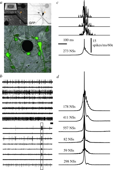
Figure 1.
The MEA and the network spike (NS). A: Cortical network on substrate-embedded multi-electrode array. The dark circle is a 30-μm-diameter electrode. Neurons are tagged using green fluorescent protein. B: Example of spontaneous activity simultaneously recorded from eight different channels. Top: at 500 s. Bottom: higher temporal resolution of 30 s from the top panel (extracted section is depicted by a dark bar). A box marks a single event of synchronous activity. C: Top three traces show examples of individual synchronous events in terms of number of spikes recorded in 60 electrodes (1 ms time bins). The average of 273 such events (NSs) is shown. D: Example of average NSs recorded over 1 h from different networks (normalized amplitudes). Figure taken by permission from reference 21.