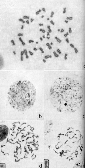
Figure 3.
Photomicrographs (× 1,200) of liver parenchymal cells from human fetuses at about fourth month of gestation. A: Female metaphase figure with 46 chromosomes; B: male interphase nucleus with a small chromocenter, presumably representing the Y, discernable at top center; C: female interphase nucleus with sex chromatin body plainly visible in lower center; D: male prophase nucleus with a small chromosome in center distinctly showing positive heteropyknosis (the inset in the lower left corner shows a small acrocentric metaphase chromosome presumed to be the Y); and E: female prophase nucleus with one large chromosome at the lower left demonstrating precocious condensation (the inset in the lower left corner shows the submediocentric metaphase chromosome presumed to be the X). (From: Ohno S, The Lancet 1961;1(7168):78, with permission from Elsevier.38)