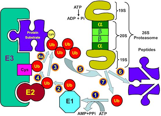
Figure 5.
The ubiquitin-proteasome proteolytic system.
Ubiquitin is activated by the ubiquitin-activating enzyme, E1 (1) followed by its transfer to a ubiquitin-carrier protein (ubiquitin-conjugating enzyme, UBC), E2 (2). E2 transfers the activated ubiquitin moieties to the protein substrate that is bound specifically to a unique ubiquitin ligase E3 (A and B). In the case of RING finger ligases, the transfer is direct (A3). Successive conjugation of ubiquitin moieties to one another generates a polyubiquitin chain (A4) that serves as the binding (A5) signal for the downstream 26S proteasome that degrades the target substrates to peptides (A6). In the case of HECT domain ligases, ubiquitin generates an additional thiol-ester intermediate on the ligase (B3) and only then is transferred to the substrate (B4). Successive conjugation of ubiquitin moieties to one another generates a polyubiquitin chain (B5) that binds to the 26S proteasome (B6) followed by degradation of the substrate to peptides (B7). Free and reusable ubiquitin is released by de-ubiquitinating enzymes (DUBs) (8).