Figure 6.
Series of interactive tools for 3D analysis are provided to quantify the patient’s condition.2
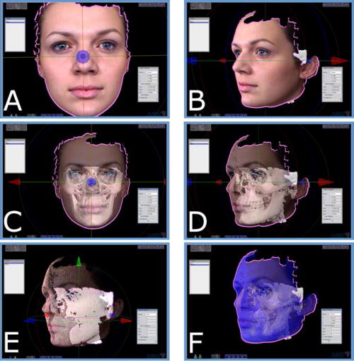
A: Surface scan image overlaid on generated skin mesh. Frontal view. B: Lateral views. C: Patient with skin surface semi-transparent to reveal underlying bone model. Frontal view. D: Lateral view. E: Wire mesh view of skin and underlying bone model. F: Wire mesh view of skin and bone model displaying underlying tie-nodes to model soft-tissue compressibility. These nodes act as springs that are differentially calibrated to represent differing tissue compressibility from the bone polygons to the nodes. Thus, this determines how the skin and bone interact.