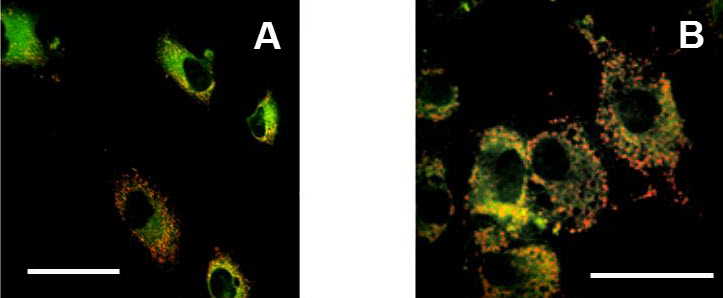
Figure 6.
Examples of microscopic images of cells stained by JC-1.
A: Control cultured osteoblasts. B: Cultured osteoblasts treated by pro-apoptotic agent (FGIN-1-27). Greater red color staining is apparent in cells treated by pro-apoptotic agent (B) in comparison to untreated osteoblasts (A), indicating a decrease in the mitochondrial membrane potential. Confocal microscopy. Scale bar 30 μm.