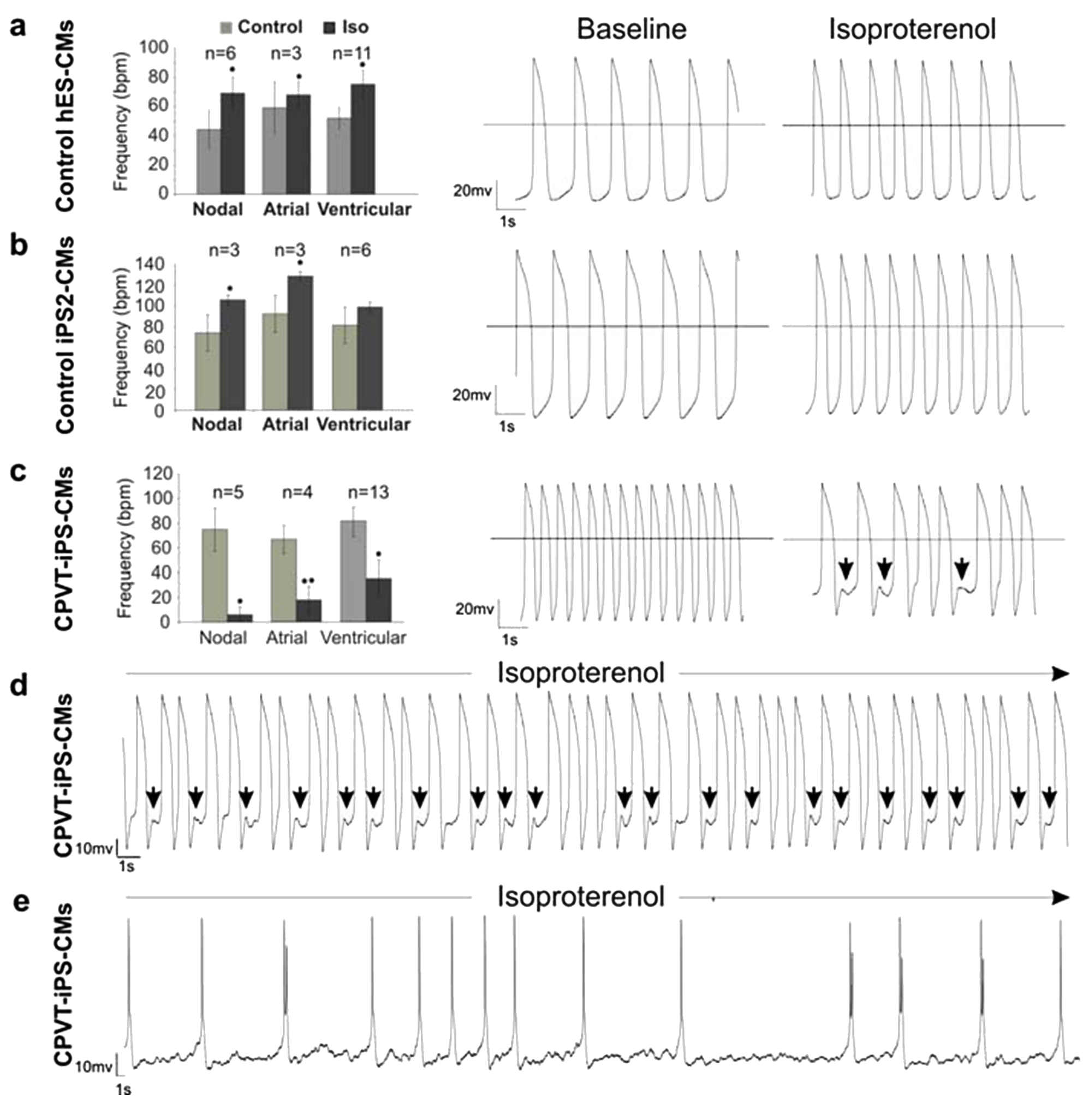
Figure 6.
Electrophysiological characterization of control and CPVT1 cardiomyocytes (CM).
Action potentials in single cardiomyocytes were recorded by the whole-cell patch clamp method in the current-clamp mode, and the beating frequency of cells at basal state and after 1 μM isoproterenol (Iso) treatment was determined. Cardiomyocytes differentiated from control embryonic stem cells (A) and iPSC lines (B) responded to 1 μM isoproterenol stimulation with positive chronotropy and did not show any arrhythmia after treatment. Representative traces of action potentials before and after isoproterenol treatment are shown in the middle and right panels, respectively. (C) A large fraction of cardiomyocytes (22 out of 38 cells, 57.9%) derived from CPVT1 iPSC responded to isoproterenol with negative chronotropy (left panel), and 13 out of 38 (34.2%) CPVT1 cardiomyocytes exhibited arrhythmia and DADs (arrows above traces in the right panel). (D,E) Representative traces of arrhythmic action potentials in two additional cells exposed to isoproterenol. The action potential traces in the cell depicted in panel (D) show putative DADs (arrows). The n values in panels (A,C) show the total number of analyzed cells. Error bars show SEM. *P < 0.05, **P < 0.01. Reprinted from Fatima A. et al.38 with permission of S. Karger AG, Medical and Scientific Publishers, Basel, Switzerland.