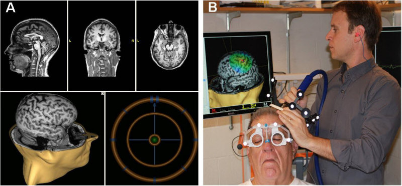
Figure 1.
The MRI-navigated Nexstim Interface.
A: Screen shot of the Nexstim neuronavigation interface: The top three panels represent the sagittal, coronal, and axial (left to right) MRI views used to locate specific spatial landmarks relative to the stereotactic spheres (shown in panel B) that are used for neuronavigated TMS. The lower panels display the stimulation target on a 3D MRI (left) and the bulls-eye target (right) that ensures that the operator holds the coil to the patient’s scalp at the correct location and orientation. B: The operator holds the figure-of-eight coil to the patient’s scalp while monitoring the brain stimulation site on the 3D MRI. Note the stereotactic spheres mounted on the coil that identify its position relative to the spheres on the patient’s goggles that localize the patient’s head.