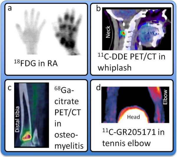
Figure 1.
Examples of PET Imaging of Peripheral Pain Mechanisms.
A:18F-FDG PET of the hand of a healthy subject and a patient with rheumatoid arthritis. Adapted from Beckers et al.9
B:11C-D-deprenyl PET/CT of a patient with whiplash-associated disorder. Adapted from Linnman et al.11
C:68Ga-citrate PET/CT of a patient affected by acute osteomyelitis of the left distal tibia. Adapted from Figure 6 (A 68Ga-citrate PET/CT scan of a patient affected by acute osteomyelitis of the left distal tibia. The scan demonstrates an area of increased tracer uptake (red area), corresponding to an area of decreased bone density on the CT images, which is consistent with acute inflammation) by Roivainen et al.16 with kind permission from Springer Science and Business Media.
D:11C-GR205171 PET of a patient with unilateral chronic tennis elbow. Adapted from Peterson.17