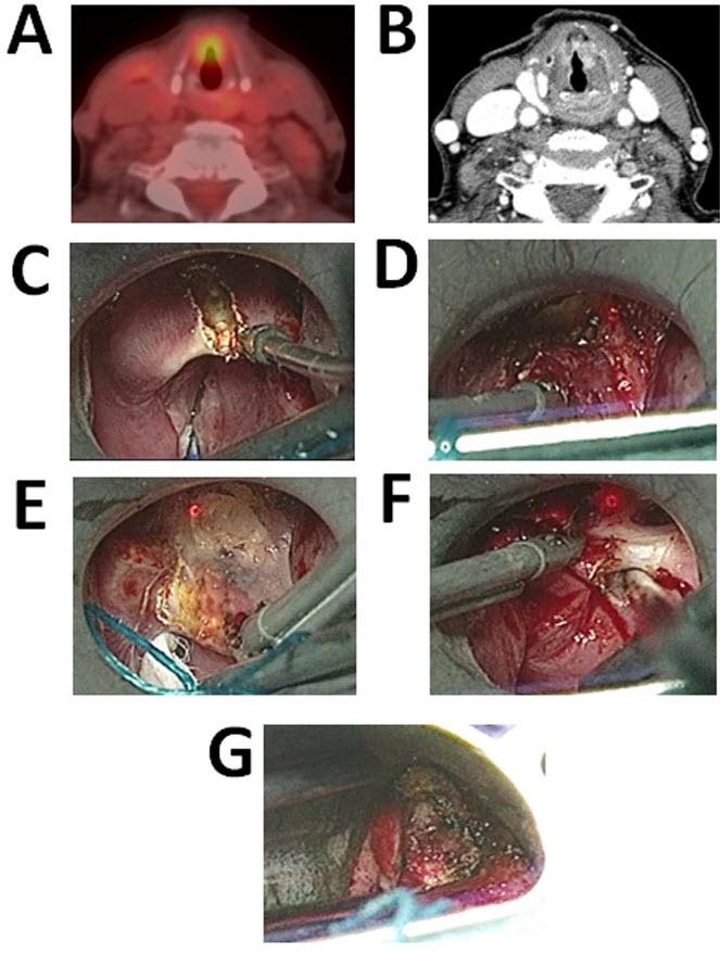
Figure 1.
Sixty-six-year-old Female with a History of Laryngeal Carcinoma Treated with EBRT and Chemotherapy Presents with Recurrent Laryngeal Cancer.
A: PET scan demonstrating FDG avid lesion of the glottis. B: Pre-operative CT scan demonstrating increased contrast enhancement at the level of the glottis. C–G: Intraoperative photographs. C: The epiglottis is divided in the midline. D: The vascular pedicle is ligated with surgical clips and divided. E: The dissection is carried out anteriorly into the pre-epiglottic space. F: The tumor is released laterally. G: Post partial laryngectomy evaluation of the glottis is performed to insure hemostasis.