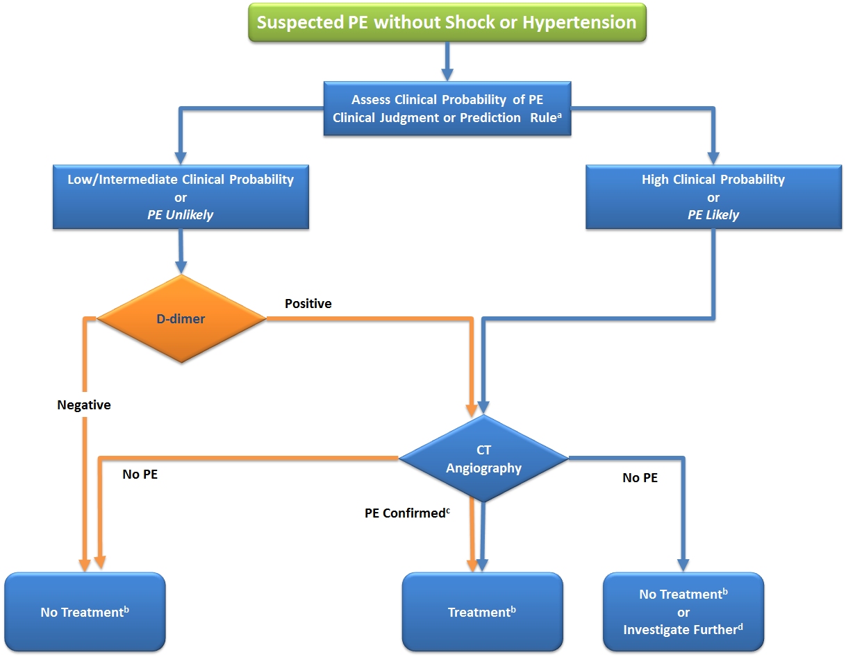
Figure 2.
Proposed Diagnostic Algorithm for Patients with Suspected High-Risk PE in the Absence of Shock or Hypotension.
a Two alternative classification schemes may be used for clinical probability assessment, i.e. a three-level scheme (clinical probability defined as low, intermediate, or high) or a two-level scheme (PE unlikely or PE likely). When using a moderately sensitive assay, D-dimer measurement should be restricted to patients with low clinical probability or a PE-unlikely classification, while highly sensitive assays may also be used in patients with intermediate clinical probability of PE due to a higher sensitivity and negative predictive value.
b Treatment refers to anticoagulation treatment for PE.
c CT angiogram is considered diagnostic of PE if it shows PE at the segmental or more proximal level.
d In case of a negative CT angiogram in patients with high clinical probability, further investigation may be considered before withholding PE-specific treatment.
CT, computed tomographic; PE, pulmonary embolism.