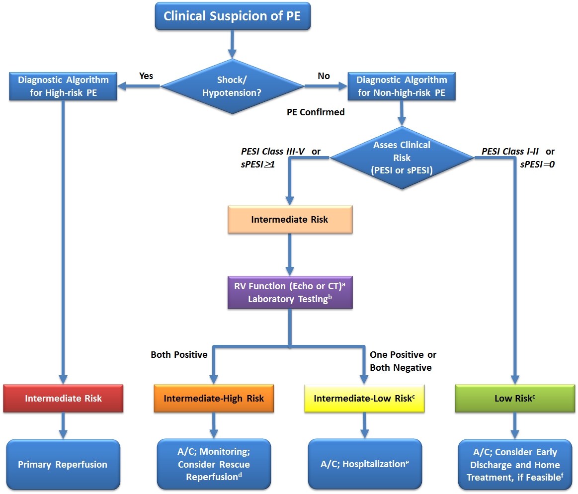
Figure 3.
Risk-Adjusted Management Strategies in Acute PE. Based on Konstantinides et al.8
a If echocardiography has already been performed during diagnostic work-up for PE and detected RV dysfunction, or if the CT already performed for diagnostic work-up has shown RV enlargement (right/left ventricular ratio ≥0.9), a cardiac troponin test should be performed except for cases in which primary reperfusion is not a therapeutic option (e.g. due to severe co-morbidity or limited life expectancy of the patient).
b Markers of myocardial injury (e.g. elevated cardiac troponin I or -T concentrations in plasma) or of heart failure as a result of (right) ventricular dysfunction (elevated natriuretic peptide concentrations in plasma). If a laboratory test for a cardiac biomarker has already been performed during initial diagnostic work-up (e.g. in the chest pain unit) and was positive, then an echocardiogram should be considered to assess RV function, or RV size should be (re)assessed on CT.
c Patients in the PESI Class I–II, or with sPESI of 0, and elevated cardiac biomarkers or signs of RV dysfunction on imaging tests are also to be classified into the intermediate-to-low-risk category. This might apply to situations in which imaging or biomarker results become available before calculation of the clinical severity index. These patients are probably not candidates for home treatment.
d Thrombolysis, if (and as soon as) clinical signs of hemodynamic decompensation appear; surgical pulmonary embolectomy or percutaneous catheter-directed treatment may be considered as alternative options to systemic thrombolysis, particularly if the bleeding risk is high.
e Monitoring should be considered for patients with confirmed PE and a positive troponin test, even if there is no evidence of RV dysfunction on echocardiography or CT.
f The simplified version of the PESI has not been validated in prospective home treatment trials; inclusion criteria other than the PESI were used in two single-armed (non-randomized) management studies.
A/C, anticoagulation; CT, computed tomographic pulmonary angiography; PE, pulmonary embolism; PESI, pulmonary embolism severity index; RV, right ventricular; sPESI, simplified pulmonary embolism severity index.