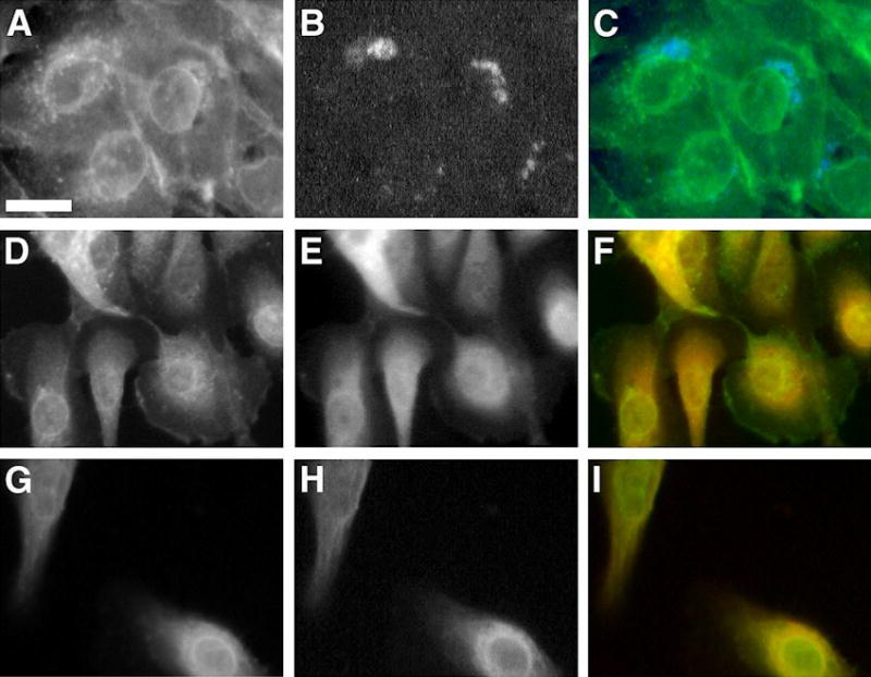
Figure 2. After Exposure to Prolactin, GLUT1 Co-localizes with ECFP-Golgi, α-Lactalbumin and α-Mannosidase II
Fluorescent images were captured 60 hours after transfection with 1 μg of pECFP-Golgi. Cells were maintained in prolactin-rich medium for 4 days, before they were fixed and stained with specific anti-GLUT1, anti-α-lactalbumin or anti-α-mannosidase II. GLUT1 is shown in green, and α-lactalbumin or α-mannosidase II in red after staining with FITC-conjugated and Texas Red-conjugated secondary antibodies, respectively. ECFP-Golgi emits cyan-blue fluorescence when exposed to fluorescent light at the appropriate wavelength. Bar 10 μm. A, D, G: GLUT1 signal. B: ECFP-Golgi signal. E, H: α-Lactalbumin and α-mannosidase II signals, respectively. C, F, I: Superimposed images. Perinuclear co-localization of GLUT1 and ECFP-Golgi is shown as areas of coincident staining (C). Co-localization of GLUT1 and α-lactalbumin or α-mannosidase II appear as areas of coincident staining, giving rise to yellow signal (F, I).