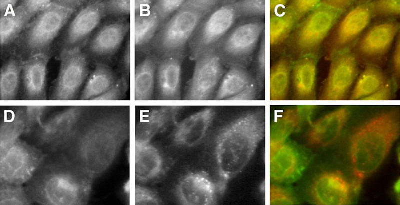
Figure 3. After Exposure to Prolactin, GLUT1 only Partially Co-localizes with β-COP and Transferrin-Texas Red in Endosomes
Cells were maintained in prolactin-rich medium for 4 days, before they were fixed and stained with specific anti-GLUT1 or anti-β-COP primary antibodies. Some cells were exposed shortly to transferrin-Texas Red staining before fixation and exposure to anti-GLUT1. GLUT1 is shown in green, and β-COP in red after staining with FITC-conjugated and Texas Red-conjugated secondary antibodies, respectively. Transferrin stain appears in red. A, D: GLUT1 signal. B, E: β-COP and transferrin (short-term exposure) signals, respectively. C, F: Superimposed images. Partial co-localization of GLUT1 with β-COP or transferrin in endosomes appears as areas of coincident staining, giving rise to yellow signal.