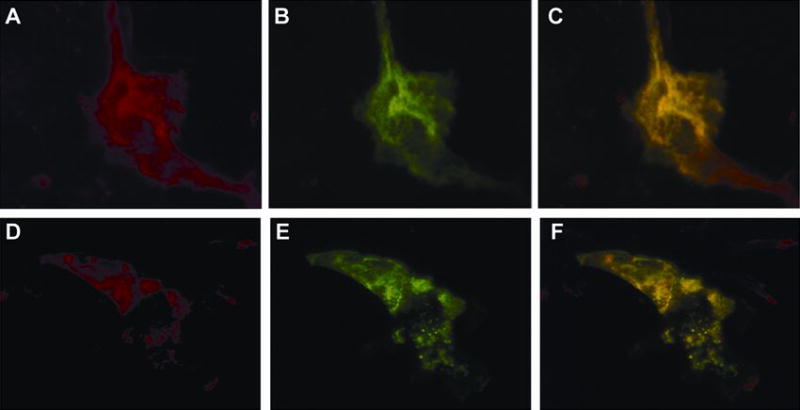
Figure 5. GLUT1 Chimeras to GFP Co-localize with Native GLUT1 in CIT3 Cells in SM
EGFP-GLUT1 fusion protein (B) exhibits the same intracellular distribution as native GLUT1 (A). Superimposed images (C) demonstrate that co-localization of native GLUT1 and EGFP-GLUT1 fusion protein appears as areas of coincident staining, giving rise to yellow signal. GLUT1-EGFP fusion protein (E) exhibits the same intracellular distribution as native GLUT1 (D). Superimposed images (F) demonstrate that co-localization of native GLUT1 and GLUT1-EGFP fusion protein appears as areas of coincident staining, giving rise to yellow signal.