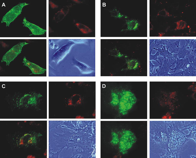
Figure 7. Degree of Differentiation Affects Intracellular Targeting of GLUT1
Static images of CIT3 cells in GM and at different levels of differentiation in SM. Panel A: In GM; B, C, D: In SM at different levels of differentiation. The upper left figure of each panel is GLUT1-EGFP signal. The upper right figure is staining of the same cells with BODIPY-TR ceramide (Molecular Probes, Inc., Eugene, OR, USA), which is a dye that marks the trans-Golgi in red. Living CIT3 cells were pre-incubated with 5 nmol/mL of BODIPY-TR ceramide at 4°C for 30 minutes. Lower left figure is superimposed image of the upper figures. Lower right figure is phase contrast image of the cells to show the different levels of differentiation.