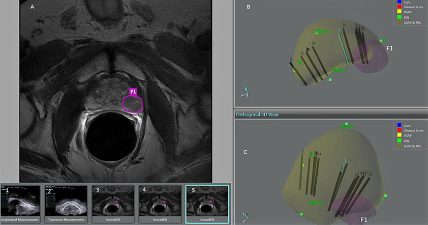
Figure 5. Multiparametric MRI-TRUS Fusion Guided Biopsy in a 62-Year-Old Male
The mpMRI-TRUS fusion guided biopsy in a 62-old year male, who had previous negative biopsy one year prior, shows a focal nodule in the left peripheral zone which underwent four core samples revealing Gleason (4+3) 7 in 80% of the specimens. The mpMRI circled region shows a left peripheral zone hypointense lesion (A). Sagittal (B) and coronal (C) depiction from 3D model of mpMRI-TRUS fusion; the left target lesion (F1) is depicted in pink. Sagittal (1) and transverse (2) TRUS images for measurement of the size of the prostate. The mpMRI images show the left peripheral zone lesion circled (3, 4, 5); these images are used to localize the F1 lesion for fusion guided biopsy.