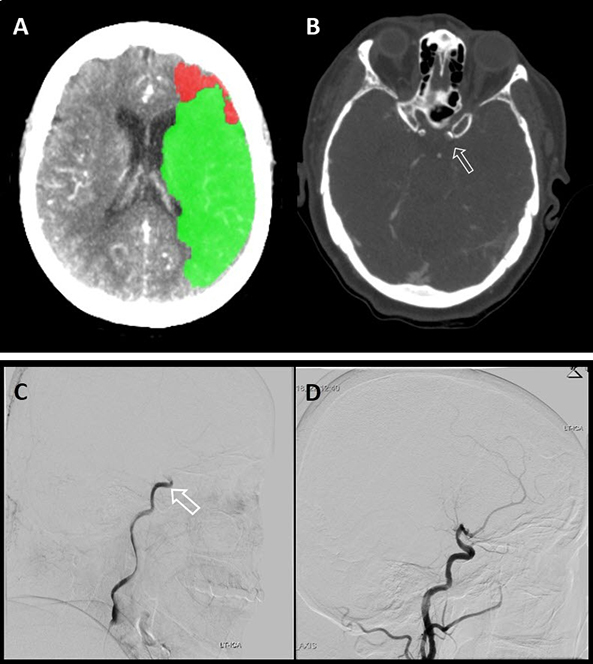
Figure 1. CT Brain Perfusion, CTA, Brain Angiography
A: A CT perfusion algorithm calculation showing decreased perfusion in a brain area consistent with left middle cerebral artery (MCA) infarct, the green area representing penumbra. B: CT angiography showing the occluded intracranial left carotid artery with a filling defect (arrow) indicating that the artery is occluded. C: Cerebral catheter angiography showing the occluded distal left carotid artery (arrow). D: Cerebral catheter angiography after successful retrieval of the clot using a stent retriever.