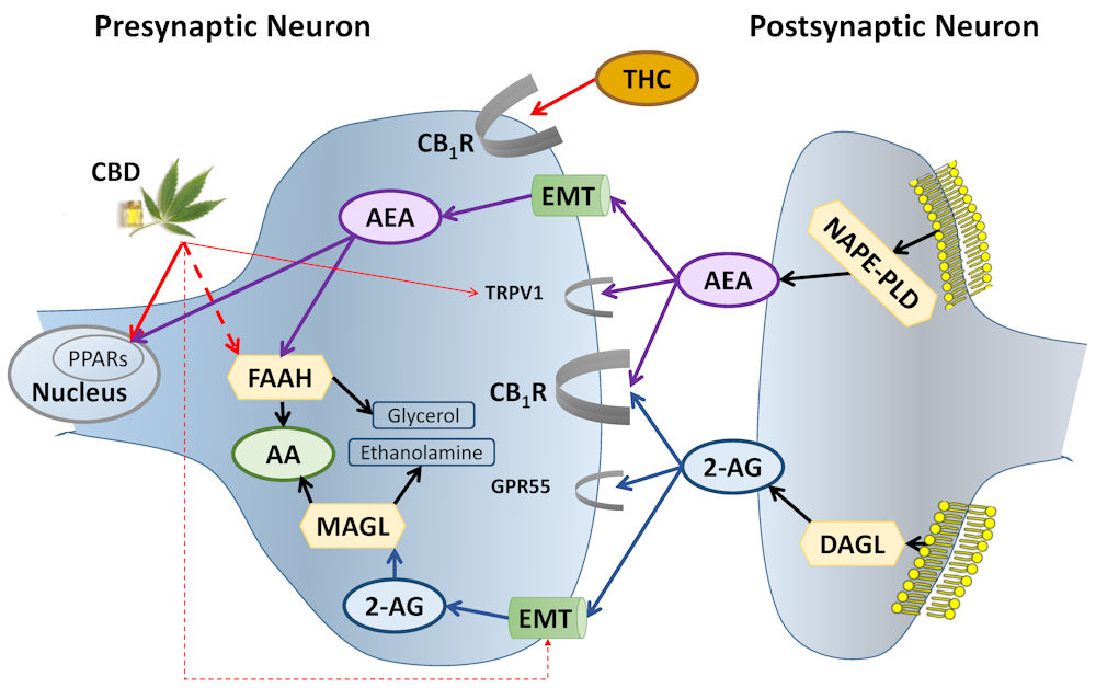
Figure 1.
Biosynthesis, Degradation, and Receptor Binding of AEA and 2-AG.
AEA is synthesized from membrane phospholipids in the postsynaptic neuron by NAPE-PLD. It crosses the synapse “against the traffic” and activates CB1R and TRPV1 on the presynaptic neuron. Following reuptake to the presynaptic neuron by the EMT, AEA activates nuclear receptors—PPARs—and is degraded by FAAH.
THC directly activates CB1R; CBD inhibits FAAH and EMT (increasing AEA levels), the endogenous ligand of CB1R. Like AEA, CBD activates PPARs and TRPV1.
2-AG, 2-arachidonoylglycerol (blue ellipses and related lines); AA, arachidonic acid (green ellipses); AEA, anandamide (purple ellipses and related lines); CB1R, cannabinoid type 1 receptor; CBD, cannabidiol; DAGL, diacylglycerol lipase; EMT, endocannabinoid membrane transporter (green tubes); FAAH, fatty acid amide hydrolase; GPR55, G protein-coupled receptor 55; MAGL, monoacylglycerol lipase; NAPE-PLD, N-acylphosphatidylethanolamine-specific phospholipase D; PPARs, peroxisome proliferator-activated receptors; THC, Δ9-tetrahydrocannabinol; TRPV1, transient receptor potential channels of vanilloid type-1. Broken lines = inhibition; half ellipses (
 ) = receptors; hexagons = enzymes; yellow mesh = cell membrane. Thicker lines and half ellipses are of greater importance than the thinner ones. Black lines, other pathways; red lines, phytocannabinoid pathways.