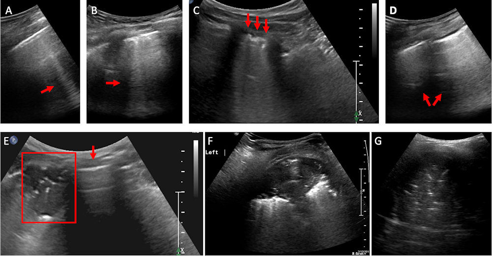
Figure 5. Ultrasound of a COVID-19 Lung Injury
A: Blurred edge B-line (red arrow). B: Multiple blurred edge B-lines (red arrow). C: Confluent blurred edge B-lines (red arrows) and thick irregular pleural line. D: Diffused B-lines “white lung” sign (red arrows). Notice the thickened pleural line. E: Lung with a small subpleural consolidation (within red frame) next to a normal, aerated lung with A-lines (red arrow). F: Subpleural consolidation with air bronchogram. G: Hepatization of the lung in segmental pneumonia with large consolidation and air bronchogram.