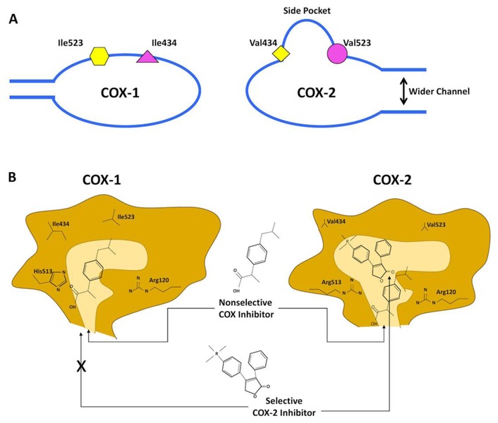
Figure 1.
Schematic Depiction of the Structural Differences between the Substrate-binding Channels of Cyclooxygenase (COX)-1 and COX-2.
Despite overall similarities, COX-1 and COX-2 show small differences in amino acid composition at the active sites. A: COX-1 has isoleucine (Ile) 523 (yellow hexagon) and 434 (pink triangle), while COX-2 has valine (Val) 523 (Val523, pink circle) and 434 (yellow square). This substitution creates a side pocket of the active binding site of COX-2, not present in COX-1. In addition, the channel of the active site in COX-2 is larger than in COX-1. Note that the non-selective COX inhibitor has access to the binding channels of both isoforms. B: The more voluminous residues (Ile523, Ile434 and Histidine (His)513) in COX-1 make a narrower entrance channel and obstruct access of the bulky side chains of the selective COX-2 inhibitor. The COX-2 side pocket allows specific binding of the selective COX-2 inhibitor side extension. Arg, Arginine. Based on Grosser et al.18