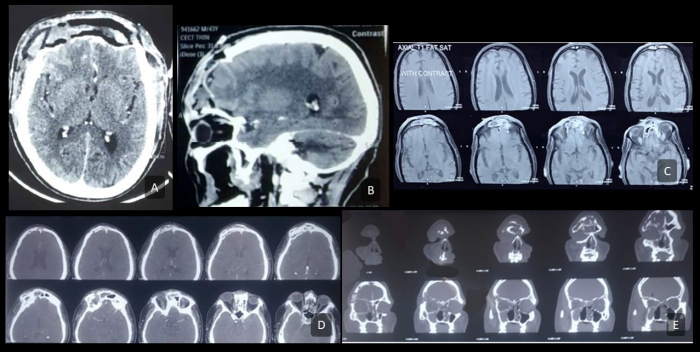
Figure 2.
Pre-operative Imaging.
A and B: Axial and sagittal contrast-enhanced CT, respectively, showing complete erosion of frontal bone with necrotic soft tissue debris between the skin and dura; the dura appears enhanced. C: T2-weighted axial MRI cuts depicting heterogeneously enhancing soft tissue density in the frontal sinus with posterior table erosion. D and E: Axial and coronal contrast-enhanced CT cuts, respectively, depicting the erosion of anterior and posterior tables of frontal sinus, suggestive of osteomyelitis.