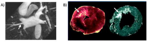
Figure 2.
Examples of arrhythmogenic anatomy depicted by MRI. A: MRI angiogram anatomy of the pulmonary veins. Note that variant pulmonary vein anatomy such as an additional right middle pulmonary vein, indicated by the white arrow, can be clearly seen by MRI. B: The complex structure of myocardial infarction scar, indicated by the white arrows, depicted by pathology on the left and delayed gadolinium enhanced MRI on the right. Figure 2A included with permission from the Journal of Cardiovascular Electrophysiology (Mansour, JCEP 2004; 15:387). Figure 2B included with permission from Circulation (Kim, Circulation 1999; 100:1992).